Rib Cage Anatomy Female / Image result for scapula and rib anatomy | Anatomy bones ... - The female's internal reproductive system is located below the kidneys.
byAdmin-
0
Rib Cage Anatomy Female / Image result for scapula and rib anatomy | Anatomy bones ... - The female's internal reproductive system is located below the kidneys.. An inhalation is accomplished when the muscular diaphragm, at the floor of the thoracic cavity, contracts and flattens, while the contraction of intercostal muscles lift the rib cage up and out. It has a roughened area on its upper surface, from which the serratus anterior muscle originates. If your specimen is female, you will find the ovaries at the base of the umbilical cord/urinary bladder. The thoracic cage takes the form of a domed bird cage with the horizontal bars formed by ribs and costal cartilages. This article will look at the osteology of the thoracic vertebrae, examining their characteristic features, joints and clinical correlations.
It is supported by the vertical sternum or. This article will look at the osteology of the thoracic vertebrae, examining their characteristic features, joints and clinical correlations. The thoracic cavity is protected by the rib cage and contains the lungs and heart. The female's internal reproductive system is located below the kidneys. This part leads to the main body of the uterus.
Human Rib Cage Spine Female Skull Clavicle and Scapula ... from atlas-content-cdn.pixelsquid.com 5th thoracic vertebra with rib. This part leads to the main body of the uterus. It has a roughened area on its upper surface, from which the serratus anterior muscle originates. The dome shaped thoracic cage provides the necessary rigidity for organ protection, weight support for the upper limbs and anchorage for muscles. The human rib cage is a component of the human respiratory system. This article will look at the osteology of the thoracic vertebrae, examining their characteristic features, joints and clinical correlations. The female's internal reproductive system is located below the kidneys. If your specimen is female, you will find the ovaries at the base of the umbilical cord/urinary bladder.
It is formed by the 12 thoracic vertebrae, 12 pairs of ribs and associated costal cartilages and the sternum.
The female's internal reproductive system is located below the kidneys. The human rib cage is a component of the human respiratory system. It encloses the thoracic cavity, which contains the lungs. An inhalation is accomplished when the muscular diaphragm, at the floor of the thoracic cavity, contracts and flattens, while the contraction of intercostal muscles lift the rib cage up and out. It forms the bony framework for breathing. It is formed by the 12 thoracic vertebrae, 12 pairs of ribs and associated costal cartilages and the sternum. If your specimen is female, you will find the ovaries at the base of the umbilical cord/urinary bladder. The thoracic cage takes the form of a domed bird cage with the horizontal bars formed by ribs and costal cartilages. This part leads to the main body of the uterus. Oct 20, 2020 · rib 2 is thinner and longer than rib 1, and has two articular facets on the head as normal. The dome shaped thoracic cage provides the necessary rigidity for organ protection, weight support for the upper limbs and anchorage for muscles. It has a roughened area on its upper surface, from which the serratus anterior muscle originates. This article will look at the osteology of the thoracic vertebrae, examining their characteristic features, joints and clinical correlations.
Jul 29, 2021 · the thoracic cage (rib cage) is the skeleton of the thoracic wall. 5th thoracic vertebra with rib. Jun 10, 2021 · the thoracic cage is a component of the thoracic wall and encloses the majority of the structures of the respiratory system. Sep 05, 2019 · along with the sternum and ribs, the thoracic spine forms part of the thoracic cage. It has a roughened area on its upper surface, from which the serratus anterior muscle originates.
Rib Cage - Medical Art Library from www.medicalartlibrary.com Bifid ribs can be seen in gorlin (nevoid basal cell carcinoma) syndrome. An inhalation is accomplished when the muscular diaphragm, at the floor of the thoracic cavity, contracts and flattens, while the contraction of intercostal muscles lift the rib cage up and out. Bifid or forked or bifurcated rib is a congenital skeletal abnormality of the rib cage with the cleaved sternal end into two. Jul 29, 2021 · the thoracic cage (rib cage) is the skeleton of the thoracic wall. It is supported by the vertical sternum or. It encloses the thoracic cavity, which contains the lungs. They are strong enough to support the skeleton and protect the vital organs in the. This part leads to the main body of the uterus.
It is formed by the 12 thoracic vertebrae, 12 pairs of ribs and associated costal cartilages and the sternum.
This article will look at the osteology of the thoracic vertebrae, examining their characteristic features, joints and clinical correlations. The thoracic cage takes the form of a domed bird cage with the horizontal bars formed by ribs and costal cartilages. 5th thoracic vertebra with rib. The human rib cage is a component of the human respiratory system. If your specimen is female, you will find the ovaries at the base of the umbilical cord/urinary bladder. Bifid or forked or bifurcated rib is a congenital skeletal abnormality of the rib cage with the cleaved sternal end into two. It has a roughened area on its upper surface, from which the serratus anterior muscle originates. An inhalation is accomplished when the muscular diaphragm, at the floor of the thoracic cavity, contracts and flattens, while the contraction of intercostal muscles lift the rib cage up and out. It forms the bony framework for breathing. It is formed by the 12 thoracic vertebrae, 12 pairs of ribs and associated costal cartilages and the sternum. The thoracic cavity is protected by the rib cage and contains the lungs and heart. Jun 10, 2021 · the thoracic cage is a component of the thoracic wall and encloses the majority of the structures of the respiratory system. The dome shaped thoracic cage provides the necessary rigidity for organ protection, weight support for the upper limbs and anchorage for muscles.
It forms the bony framework for breathing. If your specimen is female, you will find the ovaries at the base of the umbilical cord/urinary bladder. Jul 29, 2021 · the thoracic cage (rib cage) is the skeleton of the thoracic wall. It is formed by the 12 thoracic vertebrae, 12 pairs of ribs and associated costal cartilages and the sternum. Directly below the ovaries you will see a flap of tissue called the horns of the uterus;
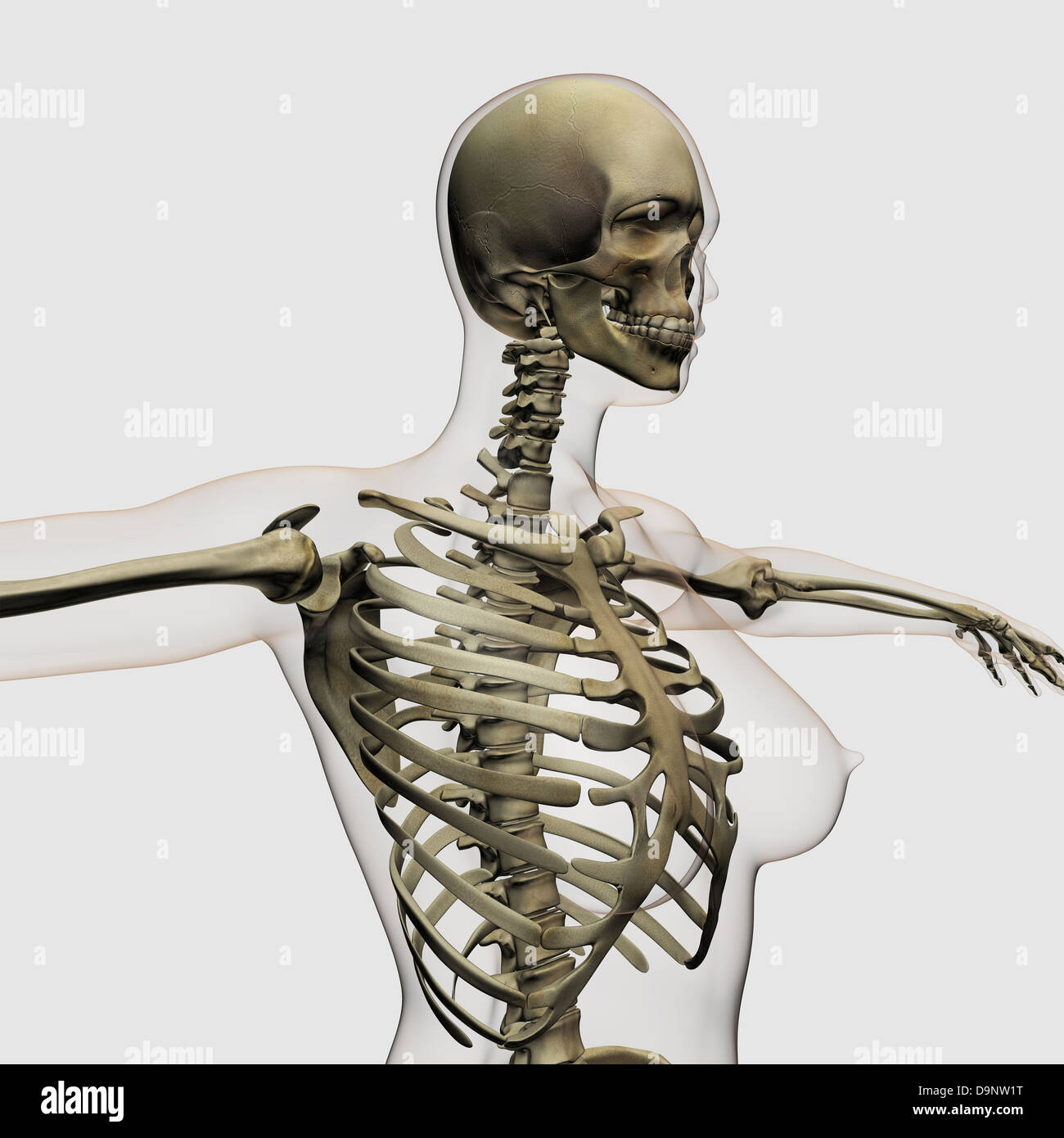
Three dimensional view of female rib cage and skeletal ... from c8.alamy.com Directly below the ovaries you will see a flap of tissue called the horns of the uterus; The human rib cage is a component of the human respiratory system. Oct 20, 2020 · rib 2 is thinner and longer than rib 1, and has two articular facets on the head as normal. Bifid or forked or bifurcated rib is a congenital skeletal abnormality of the rib cage with the cleaved sternal end into two. Jun 10, 2021 · the thoracic cage is a component of the thoracic wall and encloses the majority of the structures of the respiratory system. The thoracic cavity is protected by the rib cage and contains the lungs and heart. It is formed by the 12 thoracic vertebrae, 12 pairs of ribs and associated costal cartilages and the sternum. It encloses the thoracic cavity, which contains the lungs.
The thoracic cage takes the form of a domed bird cage with the horizontal bars formed by ribs and costal cartilages.
An inhalation is accomplished when the muscular diaphragm, at the floor of the thoracic cavity, contracts and flattens, while the contraction of intercostal muscles lift the rib cage up and out. It is supported by the vertical sternum or. It is formed by the 12 thoracic vertebrae, 12 pairs of ribs and associated costal cartilages and the sternum. 5th thoracic vertebra with rib. Bifid ribs can be seen in gorlin (nevoid basal cell carcinoma) syndrome. It encloses the thoracic cavity, which contains the lungs. Jun 10, 2021 · the thoracic cage is a component of the thoracic wall and encloses the majority of the structures of the respiratory system. This part leads to the main body of the uterus. The female's internal reproductive system is located below the kidneys. It has a roughened area on its upper surface, from which the serratus anterior muscle originates. Directly below the ovaries you will see a flap of tissue called the horns of the uterus; This article will look at the osteology of the thoracic vertebrae, examining their characteristic features, joints and clinical correlations. It forms the bony framework for breathing.
An inhalation is accomplished when the muscular diaphragm, at the floor of the thoracic cavity, contracts and flattens, while the contraction of intercostal muscles lift the rib cage up and out rib cage anatomy. Bifid ribs can be seen in gorlin (nevoid basal cell carcinoma) syndrome.